
Resource center · Early cancer diagnostics
How lateral flow assays work: strip components, formats and reading
This guide explains how lateral flow assays work: what each part of the strip does, how sandwich and competitive formats produce a line, and what affects a correct reading.
- Educational guide
- Reviewed October 2026
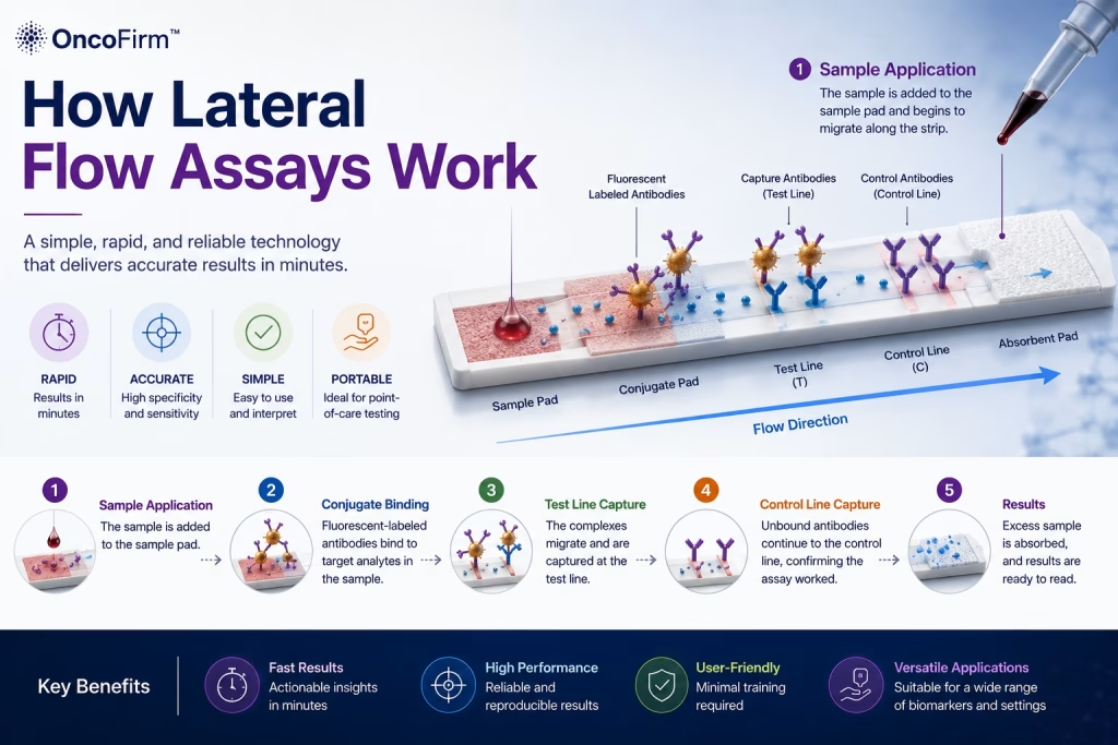
How lateral flow assays work in outline
To understand how lateral flow assays work, start with one idea. A liquid sample moves along a porous strip by capillary action and carries a labeled reagent past lines of immobilized capture reagent. Where the label is held at a line, a band forms. WHO describes malaria rapid diagnostic tests, a typical example, as lateral flow immunochromatographic tests that rely on the capture of dye-labeled antibodies to produce a visible band on a strip of nitrocellulose.
A visually read strip needs no pump, power supply or instrument, which is why the format underlies most rapid tests. The pillar guide to lateral flow tests covers uses and selection; this article covers the mechanics.
Strip components and what each one does
- Sample pad: receives the specimen and buffer and spreads the liquid evenly. It may be treated to hold back cells or adjust pH.
- Conjugate pad: holds the dried conjugate, a detection antibody or antigen coupled to a colored or fluorescent label. The sample rehydrates the conjugate and carries it forward.
- Nitrocellulose membrane: the reaction zone. Capture reagents are immobilized across it in narrow stripes that form the test and control lines.
- Absorbent pad: sits at the far end and draws liquid through the strip, which sustains flow and limits backflow.
- Backing card and housing: keep the overlapping pads in contact and frame the sample well and result window.
Many tests also use a running buffer. In WHO’s description of blood-based tests, a lysing agent ruptures the red cells and buffer carries the blood along the strip.
Sandwich format: the line appears when the analyte is present
A sandwich immunoassay uses two antibodies that bind different sites on the same target. On a strip, one antibody carries the label and the other is fixed at the test line. If the target is present it bridges the two, and label accumulates at the line. Within the working range, more analyte gives a stronger signal.
The format suits targets large enough to carry two binding sites, such as hepatitis B surface antigen, malaria antigens or hCG. Antibody tests use related layouts in which the antigen is fixed at the test line, coupled to the label, or both, so that the patient’s antibody forms the bridge.
Competitive format: the line fades when the analyte is present
Small molecules such as drugs offer only one binding site, so a sandwich cannot form. A competitive assay is used instead. Analyte in the sample competes with a labeled or immobilized version of the same molecule for a limited number of antibody binding sites.
The reading is reversed. With no analyte in the sample, label binds at the test line and a line appears. With analyte above the cut-off, binding is blocked and the line is weak or absent.
Sandwich and competitive formats compared
General behavior of the two lateral flow formats.
| Feature | Sandwich | Competitive |
|---|---|---|
| Typical analytes | Proteins and other large targets with two or more binding sites | Small molecules with a single binding site |
| Signal as analyte increases | Increases, within the working range | Decreases |
| Test line visible | Analyte detected | Analyte absent or below the cut-off |
| Test line absent | Analyte not detected | Analyte at or above the cut-off |
| Common examples | Infectious disease antigens, hCG, cardiac and tumor-associated protein markers | Drugs-of-abuse urine panels |
General principles only; the product insert defines how each test is read. Signal relationships follow Cox et al., Immunoassay Methods (Assay Guidance Manual).
The control line and reading the result
The control line captures labeled conjugate whether or not the target is present. A visible control line shows that liquid crossed the membrane and that conjugate was released. WHO cautions that it gives information on the integrity of the antibody-dye conjugate but does not confirm the ability of the test to detect the target antigen. A test with no control line is invalid and is repeated with a new device.
Results are read within the time window stated in the insert, and lines that appear later are not interpreted. Line intensity is a poor guide to quantity. WHO notes that intensity varies with the amount of antigen at least at low levels, and a laboratory study of malaria tests concluded that such tests should not be considered quantitative. Instrument reading is covered in digital cassette readers and in fluorescent vs gold nanoparticle labels.
Limits that follow from the design
- Sensitivity: a conventional strip has no amplification step, so low concentrations can be missed. For rapid influenza tests, CDC advises that negative results do not exclude infection.
- High-dose hook (prozone) effect: in sandwich tests, a very high antigen concentration can weaken the test line. The malaria study cited below reproduced the effect, although without false-negative results.
- Specificity: antibodies can cross-react with related molecules, and some specimens contain interfering substances.
- Prevalence: CDC notes that positive and negative predictive values vary considerably with how common the condition is in the population tested.
- Handling: the wrong specimen type or volume, poor storage, expired kits and late reading all affect results.
These limits are why a lateral flow result is an aid to diagnosis, interpreted by a clinician with other findings, and why the product insert is the reference for each test.
Where First Diagnostic fits
First Diagnostic™ Corporation brands and distributes lateral flow rapid tests made by established IVD manufacturers, for professional in vitro diagnostic use. The catalog includes qualitative PSA, CEA and AFP tests in the tumor-marker and general health range; these are aids used alongside other clinical and laboratory findings, not cancer screening or diagnostic tools. Rapid cancer-marker testing is a longer-term interest for the company, with nothing further available yet; contact the sales team for current products.
Frequently asked questions
What does the control line prove?
It shows that liquid migrated along the membrane and that labeled conjugate was released. It does not prove that the test line can detect the target, which is why external quality control materials are also used.
Why can a very strong positive sample give a weak line?
In a sandwich test, a very high antigen concentration can occupy the labeled antibody and the capture antibody separately, so fewer complete sandwiches form at the test line. This is the high-dose hook, or prozone, effect.
Can a lateral flow assay give a quantitative result?
A visually read test is qualitative. Some systems pair the cassette with a calibrated reader to report a numeric value, but only where the product is designed and labeled for that use.
Keep reading
- Fluorescent vs gold nanoparticle labels — How the label affects reading method, sensitivity and equipment.
- Digital cassette readers for lateral flow tests — What instrument reading adds to a visually read cassette.
- What are rapid diagnostic tests (RDTs)? — Definitions, formats and typical uses.
- Point-of-care testing: where rapid tests fit — Using rapid tests near the patient.
- Lateral flow tests explained — The pillar guide: what lateral flow tests are, where they are used and how to choose them.
References
- World Health Organization, Global Malaria Programme. How malaria RDTs work. Accessed October 2026. who.int
- Cox KL, Devanarayan V, Kriauciunas A, et al. Immunoassay Methods. In: Markossian S, Grossman A, Baskir H, et al., editors. Assay Guidance Manual. Bethesda (MD): Eli Lilly & Company and the National Center for Advancing Translational Sciences; 2012 (updated 2019). NCBI Bookshelf. ncbi.nlm.nih.gov
- Luchavez J, Baker J, Alcantara S, et al. Laboratory demonstration of a prozone-like effect in HRP2-detecting malaria rapid diagnostic tests: implications for clinical management. Malaria Journal. 2011. doi:10.1186/1475-2875-10-286. PubMed PMID 21957869. pubmed.ncbi.nlm.nih.gov
- Centers for Disease Control and Prevention. Rapid Influenza Diagnostic Tests. Accessed October 2026. cdc.gov
- Centers for Disease Control and Prevention. Rapid Diagnostic Testing for Influenza: Information for Clinical Laboratory Directors. Accessed October 2026. cdc.gov
Source: First Diagnostic™ editorial team; sources listed above · Last reviewed: October 2026 · First Diagnostic™ editorial team
Regulatory status: Products shown on this website are for professional in vitro diagnostic use. They are not cleared, approved or authorized by the U.S. Food and Drug Administration and are not offered for sale in the United States. Product availability and regulatory status vary by country; contact us for the status in your market.