
Resource center · Early cancer diagnostics
Tumor antigen detection: markers, immunoassays and limits
Tumor antigen detection means measuring molecules associated with tumors, usually proteins, with antibody-based tests. This guide covers the main antigen classes, the methods used and why a result is an aid, not a diagnosis.
- Educational guide
- Reviewed October 2026
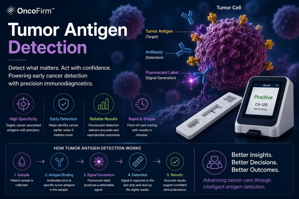
What tumor antigen detection means
Tumor antigen detection is the measurement of molecules associated with tumors, most often proteins, using antibodies that bind them. An antigen is any molecule that an antibody or immune cell can recognize. A tumor antigen is one produced by tumor cells.
The term is used in two fields. Immunologists use it for targets that the immune system, or an immunotherapy, can recognize on cancer cells. Laboratories use it more loosely for the protein tumor markers measured in blood or tissue. The U.S. National Cancer Institute (NCI) defines a tumor marker as anything present in or produced by cancer cells, or by other cells of the body in response to cancer or certain benign conditions, that provides information about a cancer. This article takes the laboratory view; the wider picture is in what are cancer biomarkers.
Classes of tumor antigen
Immunology textbooks group tumor antigens by origin:
- Mutated proteins: point mutations or gene rearrangements create new epitopes found only on the tumor. These tumor-specific antigens are often called neoantigens.
- Differentiation antigens: proteins of the tissue from which the tumor arose, such as tyrosinase in melanoma.
- Overexpressed antigens: normal proteins present at abnormally high levels on tumor cells, such as HER2.
- Abnormally modified antigens: for example, underglycosylated forms of the mucin MUC1.
- Cancer-testis antigens: proteins normally expressed only in male germ cells, such as the MAGE family.
- Oncoviral proteins: viral proteins involved in the cancer process, such as HPV E6 and E7.
Most antigens measured in blood are tumor-associated, not tumor-specific: normal tissue makes them too. PSA is produced by normal as well as malignant prostate cells. CEA and AFP are often described as oncofetal antigens, proteins made mainly during fetal development that can reappear at raised levels in some cancers and in some benign conditions. This shared origin is the root of their limited specificity.
Methods used for tumor antigen detection
Almost all methods are immunoassays. For soluble antigens the usual design is a sandwich, in which two antibodies bind different sites on the antigen and the signal rises with the amount present. The methods differ in how the signal is generated and read, and in whether the result is a number or a yes-or-no answer.
Immunoassay methods compared
Common methods for measuring protein tumor antigens.
| Method | Specimen | Result | Typical setting |
|---|---|---|---|
| Automated laboratory immunoassay (for example chemiluminescent) | Serum or plasma | Quantitative | Hospital and reference laboratories; serial monitoring |
| ELISA (microplate) | Serum or plasma | Quantitative | Laboratories running samples in batches |
| Immunohistochemistry | Tumor tissue section | Staining pattern scored by a pathologist | Pathology laboratories; treatment selection |
| Lateral flow test | Whole blood, serum or plasma, depending on the product | Qualitative against a cut-off; numeric with some reader-based systems | Near-patient professional use |
General characteristics. Specimen types and intended use are defined by each product’s instructions for use.
What a tumor antigen result is used for
NCI lists the established uses of tumor markers: helping to diagnose cancer together with other tests, determining type and stage, estimating prognosis, selecting treatment, monitoring response and checking for recurrence.
Monitoring relies on serial measurements, where the trend matters more than any single value. CEA, for instance, is used to follow how well treatment is working and whether colorectal cancer has returned. HER2 and PD-L1 are assessed in tumor tissue to help determine treatment.
Limits of tumor antigen testing
Three limits apply to nearly every circulating tumor antigen, and NCI states them plainly:
- An elevated level does not mean that a person has cancer, because noncancerous conditions can raise it. NCI’s evidence summary on ovarian cancer screening, for example, notes raised CA-125 in endometriosis, liver disease and congestive heart failure.
- A normal level does not exclude cancer. Not everyone with a given cancer has a raised level of the associated marker.
- Circulating markers have generally not worked well for screening people without symptoms, for both of the reasons above.
Diagnosis therefore rests on clinical assessment, imaging and pathology, with marker results as supporting evidence. The evidence standards for testing people without symptoms are discussed in why early detection matters.
Analytical points for laboratories
Results for the same antigen can differ between assay methods, so serial monitoring is normally done with one method and reported against that method’s reference range. Antibodies present in some patients’ samples can also interfere with immunoassays.
Sandwich assays, including lateral flow tests, can show a high-dose hook effect, in which a very high antigen concentration gives a weaker signal than expected. A qualitative strip reports only whether the antigen is above its cut-off, and line intensity is not a measurement. The mechanics are explained in how lateral flow assays work, and instrument-read labels in fluorescent vs gold nanoparticle labels.
Where First Diagnostic fits
First Diagnostic™ Corporation distributes rapid tests for professional in vitro diagnostic use, including qualitative PSA, CEA and AFP tests in its tumor-marker and general health range. These are aids used alongside other clinical and laboratory findings, not cancer screening or diagnostic tools. Rapid cancer-marker testing is a longer-term interest for the company, with nothing beyond the current catalog available yet. For inserts and regulatory documentation, contact the sales team.
Frequently asked questions
Is a tumor antigen the same as a tumor marker?
The terms overlap. A tumor antigen is a molecule from a tumor that antibodies or immune cells can recognize. A tumor marker is anything that gives information about a cancer, including proteins, gene changes and other substances. Protein markers such as PSA, CEA and AFP are both.
Does an elevated tumor antigen level mean cancer?
No. Benign conditions can raise the level of most circulating tumor antigens, and a raised result needs clinical assessment and further investigation. A normal level does not exclude cancer either.
Why are most tumor antigens unsuitable for screening?
Because they are made by normal tissue as well as by tumors, and because not all tumors release them in measurable amounts. NCI states that circulating tumor markers have generally not been sensitive or specific enough to work well for screening.
What does a qualitative rapid test for a tumor antigen show?
It shows whether the antigen in the specimen is above or below the cut-off defined in the product insert. It does not give a concentration, and the result is read and interpreted by a professional according to the insert, together with other clinical and laboratory findings.
Keep reading
- What are cancer biomarkers? — Definitions, types by clinical purpose and what a marker result can show.
- Why early detection matters — Early diagnosis versus screening, and what the evidence supports.
- How lateral flow assays work — Strip components, sandwich and competitive formats, and reading results.
- Fluorescent vs gold nanoparticle labels — How the label affects reading method, sensitivity and equipment.
- Lateral flow tests explained — The pillar guide: what lateral flow tests are, where they are used and how to choose them.
References
- National Cancer Institute. Tumor Markers. Reviewed December 7, 2023. cancer.gov
- National Cancer Institute. Tumor Marker Tests in Common Use. Reviewed December 7, 2023. cancer.gov
- Janeway CA Jr, Travers P, Walport M, et al. Immunobiology: The Immune System in Health and Disease. 5th edition. New York: Garland Science; 2001. Section 14-12, T lymphocytes can recognize specific antigens on human tumors. NCBI Bookshelf. ncbi.nlm.nih.gov
- National Cancer Institute. Prostate-Specific Antigen (PSA) Test. Reviewed January 31, 2025. cancer.gov
- National Cancer Institute. Ovarian, Fallopian Tube, and Primary Peritoneal Cancers Screening (PDQ®)–Health Professional Version. Updated April 9, 2025. cancer.gov
- Cox KL, Devanarayan V, Kriauciunas A, et al. Immunoassay Methods. In: Markossian S, Grossman A, Baskir H, et al., editors. Assay Guidance Manual. Bethesda (MD): Eli Lilly & Company and the National Center for Advancing Translational Sciences; 2012 (updated 2019). NCBI Bookshelf. ncbi.nlm.nih.gov
- Luchavez J, Baker J, Alcantara S, et al. Laboratory demonstration of a prozone-like effect in HRP2-detecting malaria rapid diagnostic tests: implications for clinical management. Malaria Journal. 2011. doi:10.1186/1475-2875-10-286. PubMed PMID 21957869. pubmed.ncbi.nlm.nih.gov
Source: First Diagnostic™ editorial team; sources listed above · Last reviewed: October 2026 · First Diagnostic™ editorial team
Regulatory status: Products shown on this website are for professional in vitro diagnostic use. They are not cleared, approved or authorized by the U.S. Food and Drug Administration and are not offered for sale in the United States. Product availability and regulatory status vary by country; contact us for the status in your market.