
Guide
Lateral flow tests explained
Lateral flow tests are the technology behind most rapid tests. This guide covers how the strip works, the main test formats, how results are read and where the limits are.
- Guide for professionals
- Reviewed October 2026
What lateral flow tests are
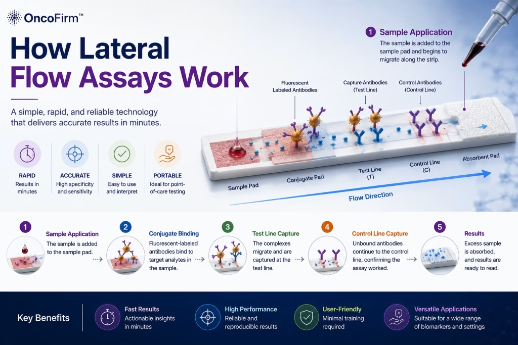
A lateral flow test, also called a lateral flow assay or immunochromatographic test, is a paper-based device that detects a target substance in a liquid specimen and shows the result as colored lines. The specimen moves along the strip by capillary action; no pump, power or instrument is needed.
Most rapid tests for infectious disease use this format, because it is quick to run, stable at room temperature and readable by eye.
The parts of a lateral flow test
- Sample pad. Receives the specimen, with or without a buffer, and filters it.
- Conjugate pad. Holds dried antibodies or antigens attached to colored particles, commonly colloidal gold. The specimen rehydrates them.
- Membrane. A nitrocellulose strip carrying two reagent lines: the test line, which captures the target, and the control line, which captures excess conjugate.
- Absorbent pad. Draws the liquid through the strip.
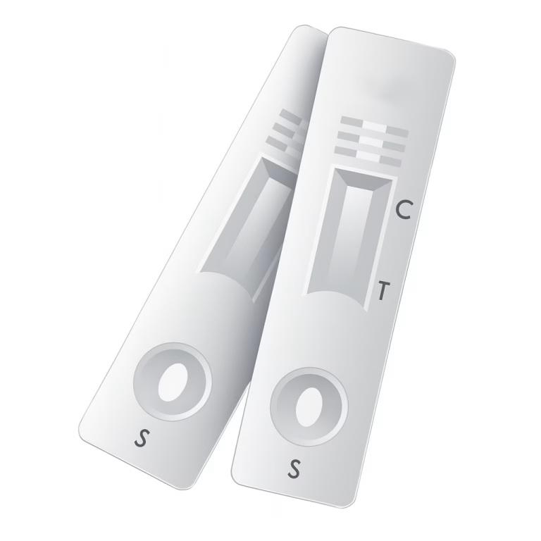
- Cassette. The plastic housing with a sample well and a result window marked T (test) and C (control).
A more detailed walk-through is in how lateral flow assays work.
Antigen tests and antibody tests
The two formats answer different questions. Examples are from the First Diagnostic range.
| Antigen test | Antibody test | |
|---|---|---|
| What it detects | Part of the organism itself | The patient’s immune response to the organism |
| What a positive suggests | Antigen from the organism was detected; some antigens remain detectable for a time after treatment | The patient has been exposed; the infection may be current or past |
| Typical specimen | Swab, urine, whole blood | Whole blood, serum, plasma |
| Main limitation | Low organism numbers can give a negative result | Antibodies take time to appear and can persist after cure |
| Examples | Chlamydia, gonorrhea, malaria P.f., influenza A & B, HBsAg | Syphilis, HCV, dengue IgG/IgM, rubella IgG/IgM |
How a lateral flow test is run
The exact procedure is defined by each product insert. In outline:
- Prepare. Bring the sealed pouch and the specimen to room temperature. Check the expiry date and open the pouch just before use.
- Add the specimen. Apply the stated volume to the sample well. Swab and urine tests need an extraction step first.
- Add buffer if required. Whole blood tests usually need drops of buffer to carry the specimen along the strip.
- Wait for the read time. Start a timer. Read at the time stated in the insert, not earlier.
- Read and record. Check the control line first, then the test line or lines. Record the result with the lot number and discard the device as biohazardous waste.
Always follow the instructions for use supplied with the kit.
Reading the lines
- Control line. It must appear. It shows that enough liquid flowed along the strip. No control line means the result is invalid and the test is repeated with a new device.
- Test line. In the sandwich format used by most tests in our range, a line at T means the target was detected. A faint line is still a line, unless the insert says otherwise.
- Read window. Each insert gives a time to read and a time after which the result must not be interpreted, because lines can appear or fade as the strip dries.
- Multi-line tests. Combination cassettes carry several test lines, for example T1, T2 and T3 on the HIV / HBsAg / HCV combo. Read each line against the insert.
What lateral flow tests can and cannot tell you
A lateral flow test gives a qualitative answer: detected or not detected at the level the test can see. It does not measure how much of the target is present, and it does not replace laboratory methods where those are required.
- A negative result does not exclude infection. The specimen may have been taken too early, or the target may be below the detection level.
- A positive antibody result does not always mean active disease.
- How often a positive result is true depends on how common the condition is in the people tested.
- Results are an aid to diagnosis and are interpreted by a clinician with other clinical and laboratory findings.
Good practice that protects results
- Store kits within the temperature range on the label and never freeze cassettes.
- Use the dropper, swab and buffer supplied with the kit.
- Train every operator on the insert, and repeat training when a new lot or product arrives.
- Run external positive and negative controls as your quality system requires.
- Keep lot numbers with results so that a problem can be traced.
Our article on point-of-care testing covers quality practice in more detail, and digital cassette readers looks at instrument-assisted reading.
Lateral flow tests in our range
Chlamydia and gonorrhea antigen, syphilis antibody.
HBsAg, HCV and combination cassettes.
Antigen and antibody cassettes for whole blood.
Influenza A and B antigen from swab specimens.
Frequently asked questions
Is a lateral flow test the same as a rapid test?
Most rapid tests are lateral flow tests, but not all. Reagent strips for urinalysis, for example, are rapid tests that work by a color reaction on a pad rather than by lateral flow.
How long does a lateral flow test take?
The tests with published inserts are read between 5 and 20 minutes after the specimen is added. The exact time and the read window are stated in each insert.
Does a faint test line count?
For the sandwich-format tests in our range, the inserts say that any visible test line within the read window is read as positive. Follow the insert for the product you are using.
Can lateral flow results be read by an instrument?
Yes, some tests are designed for reader instruments. A First Diagnostic cassette reader is a development goal and is not available to order.
Keep reading
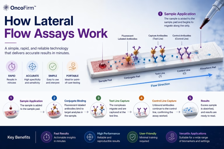
A technical guide to the lateral flow strip: what each component does, how sandwich and competitive formats produce a line, and what limits a correct reading.
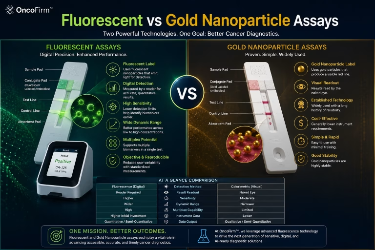
Colloidal gold gives a line read by eye; fluorescent labels need a reader. This guide compares the two for sensitivity, quantitation, equipment and workflow.
What rapid diagnostic tests are, the formats and analyte types in use, where they fall short of laboratory methods, and five points to check before buying an RDT.
Source: First Diagnostic™ editorial team; product inserts for the products named; sources listed above · Last reviewed: October 2026 · First Diagnostic™ editorial team
Regulatory status: Products shown on this website are for professional in vitro diagnostic use. They are not cleared, approved or authorized by the U.S. Food and Drug Administration and are not offered for sale in the United States. Product availability and regulatory status vary by country; contact us for the status in your market.